4 May 2020

New research led by scientists at the Australian Regenerative Medicine Institute (ARMI) has unveiled how white blood cells move and carry out their functions at the cellular and subcellular level – at a resolution at which we have not seen before.
PhD student Harriet Manley spearheaded the work with ARMI Group leader Professor Graham Lieschke which was recently published in the Journal of Leukocyte Biology. Collaborators included researchers from Monash Micro Imaging (MMI) and the Advanced Imaging Center at the HHMI Janelia Research Campus (USA).
“The movement of white blood cells underpins the inflammatory response – how our immune system responds to insults, injuries and infections,” explained Harriet. “The challenge in studying white blood cell behaviour within living organisms is that to be effective, cells in the immune system must move and change shape incredibly quickly while travelling through the bloodstream or squeezing through tight spots. Imaging technology, up until now, has struggled to meet this challenge.”
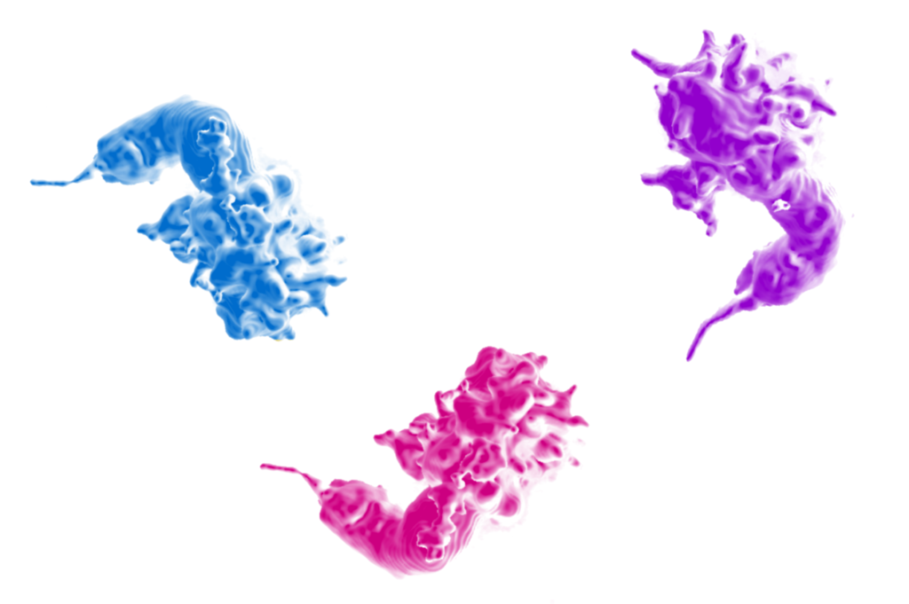
A single zebrafish neutrophil captured at successive timepoints by intravital lattice lightsheet microscopy (pseduo-coloured 3-dimensional surface render images). Lattice lightsheet microscopy provides vastly improved spatiotemporal resolution, enabling rapid leukocyte dynamics and fine details of leukocyte morphology to be visualized in their native physiological setting.
Using cutting-edge lattice lightsheet microscopy (LLSM), the research team have been able to image white blood cells in live zebrafish at amazing spatial and temporal resolution. This work contributes immensely to our understanding of white blood cells. The findings show how white blood cells change their morphology (i.e. shape) during migration, and reveal interactions between white blood cells and blood vessel cells that have never been observed, and new details on white blood cell structures and behaviours.
This research led to the first publication using Monash University’s 3i lattice lightsheet microscope housed at MMI. Harriet added, “David Potter from MMI and I were the pioneering partnership to get this Lieschke Group project going at Monash, a project which was then propelled by Graham and I visiting the Advanced Imaging Center at Janelia Research Campus in April 2018. Over in the US, we received invaluable assistance from our collaborators, spending 12 days of unlimited imaging using their lattice lightsheet microscope to generate a whopping 30 terabytes of data! Data storage and management was also made possible by Monash’s MASSIVE team.”
This powerful and advanced imaging technology is set to revolutionise biomedical research. Considering the currently-topical research interest in inflammation and immunomodulation, such as immunotherapy for a range of diseases, including many cancers, a better understanding of the underlying biology of white blood cells is of paramount importance. This is the new benchmark in white blood cell imaging within living organisms. This LLSM of white blood cells sets the stage for transformative studies into the cellular and subcellular complexities of their biology.
Read the full media release.